Applications of Accelerators in Nuclear Science from the Viewpoint of Photon Activation Analysis
Philip Cole, Physics Dept. Lamar University
 I would first like to thank the organizers of the Forum on International Physics for the opportunity to give an invited talk on the Applications of Accelerators in Nuclear Science from the Viewpoint of Photon Activation Analysis in the section Opportunities in Global Science Industries for the 2018 APS April Meeting. Not only was it a joy to give the talk, but it serendipitously resulted in a book contract. A representative of Cambridge University Press was in the audience. The upcoming second edition1 will be a thoroughly revised and updated edition of Photon Activation Analysis2, which is currently out of print.
I will give a brief background on the physics of photon activation. Photon Activation Analysis (PAA) is based upon the photonuclear interaction. When a photon of energy 15 MeV, say, strikes a nucleus, a neutron can be liberated, and through these means, an excited, proton-rich nuclide is formed. These photons are produced, for example, when a 30-MeV electron beam is directed onto a rather thick slab of a high-Z radiator, usually of thickness of around two radiation lengths. Most of the post-bremsstrahlung electrons will be stopped in this thick radiator or converter. The bremsstrahlung photons have a characteristic 1/E fall-off and will span the entire cross section of the giant dipole resonance curve. Thus, the yield of knockout neutrons is a convolution of the bremsstrahlung spectrum with the photonuclear cross section in the giant dipole resonance regime as can be seen in Fig. 1.
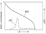
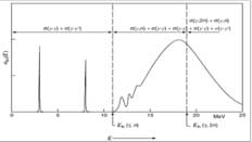
Fig. 1. (Left) Bremsstrahlung continuum and photonuclear cross section; Φ(E), differential bremsstrahlung photon flux density; and σ(E), differential cross section, both as functions of photon energy. (Right) Total (γ,γ’) and (γ, xn) absorption cross section; σtot, total photon absorption cross section; Eth, threshold energy.
During photon activation analysis, nuclides of the analyte elements in the material samples are excited to radioactive nuclides. The high-energy photon interacts with the target nucleus and in the ensuing photonuclear reaction, a nucleon (proton or neutron) is ejected from the probed nucleus. Usually this nucleon is a neutron, as the Coulomb energy barrier will tend to inhibit protons from escaping. In fact, the most important contribution to the total photoneutron cross section in the giant dipole resonance region, by far, is single-neutron emission from the photo-excited nucleus. The resulting nuclide will be a proton-rich isotope of that interrogated element. In most cases, the isotope is unstable and this excited nuclide will cascade down to its ground state; usually through emitting several gamma rays, each having a characteristic energy ranging from ~100 keV to several MeV. Measuring these discrete gamma rays will “fingerprint” the nuclide; the resulting characteristic spectral lines can then be identified with an appropriate spectrometer, such as a high-purity Germanium (HPGe) detector as indicated in Fig. 2. And through the spectroscopy of identifying peaks within the complete gamma-energy spectrum, each chemical element – not the chemical species – can be detected qualitatively.
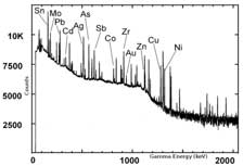
Fig 2. A typical gamma energy spectrum of multi-element sample, where the elements can be identified by the characteristic gamma energies.
Photon activation analysis gives information on the type of element present within the sample. It cannot, however, determine the absolute number of nuclides activated. One cannot know a priori the exact mass of each of the separate elements within a random sample. Assuming uniform irradiation of the sample under interrogation, with all other beam parameters held fixed, the number of the proton-rich nuclides produced will scale with the absolute number of the interrogated isotopes of a specific element. Once the photon activation period is complete, the decay gamma rays from the excited nuclides will be measured. And similarly a greater number of the photoproduced nuclides will be reflected by the higher intensities of the characteristic spectral signals, which are emitted from the proton-rich nuclides as they decay to ground state. To determine the absolute masses of specific elements of the samples under scrutiny, a calibration material will have to be irradiated along with the interrogated sample. One usually employs a standard calibration material, where the elemental components are well known. After making the necessary spectroscopic measurements, we compare the spectra of the sample with that of the calibration material. We then can exactly determine the elemental make-up and thereby can ascertain the absolute concentration of specific elements present within the sample.
The primary advantage of this in situ method of PAA (sample with a reference or calibrator) is that it can be carried out non-destructively. One does not need to specially prepare or machine the sample and hence there is a reduced danger of contamination from the debris of aggregated particles. And with this ease of handling, many more kinds of materials are open to investigation, especially materials, which are difficult to treat chemically, such as dust particles, fly ash, etc. Another advantage of PAA is its inherent high sensitivity. PAA has wide latitude of applications, ranging from the very small, i.e. minute mass, such as microgram dust particles, to the very big, i.e. massive multi-kilogram samples. In any case, should one wish to element analyze a large object – be it precious or inexpensive – large volume irradiation facilities can be engineered for employing PAA. This is easily achieved by using an electron accelerator with a suitably designed bremsstrahlung converter which will allow for large-aperture rastering to provide an appropriate photon beam flux for proper activation.
In the talk I touched upon several applications:
- environmental (dust particles, volcanic ash, electronic waste, etc.),
- agriculture (e.g. coffee plant provenance and the soil),
- biological (plant materials and tissues),
- forensic science,
- geological and extraterrestrial materials (ores, meteorites, lunar samples, etc.),
- art and archaeology,
- raw products, industrial, and high purity material,
- waste material assay, and
- Harvey recovery assay. [this tropical storm dumped 1.2 m of rain in SE Texas over 4 days]
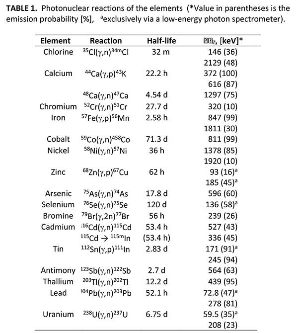
Tabulated in Table 1 below, we find the photonuclear reactions, where the second column expresses the primary one-neutron knockout (see above) mechanism for photoproducing a nuclide or isomer. An excited nuclide will tend to beta decay and subsequently give off specific γ rays (photons) as it goes to ground state; these gamma-energy fingerprints are delineated in the fourth column. These spectral lines (γ rays) can be identified from a well-calibrated HPGe detector described above. In the third column one can see the half-life or the time for half the excited nuclides to decay. For the elements of interest, the decays can be mere seconds to days. This process follows an exponential-decay curve.
For low-Z elements, from H to B, no analytically useful radionuclides are produced. From carbon, nitrogen, oxygen and fluorine pure β+-emitters are produced by (γ,n)-reactions. These radionuclides cannot be analyzed by means of nuclide specific gamma-rays, but only by detecting the unspecific 511-keV annihilation radiation, which originates from positrons emitted from radioactive nuclei being absorbed in the surrounding material. In the annihilation process, a positron captures an electron from the material forming positronium, which then decays by conversion of the positron and electron rest mass (511 keV each) into two 511-keV photons. These two 511-keV photons are simultaneously emitted into opposite directions. Since only the 511-keV annihilation radiation is available for the analysis of the light elements up to fluorine, a chemical separation of these elements must be performed.
In the case of medium and heavy elements, however, the radionuclides produced by photonuclear reactions emit – with very few exceptions – characteristic gamma-radiation. Due to the specific gamma-radiation an instrumental multi-element analysis of medium and heavy elements using high resolution gamma-ray spectroscopy becomes possible. In general, photoneutron reactions – (γ,n), (γ,2n), etc. – result in neutron-deficient nuclides. In the case of low-atomic-number nuclei, their predominant decay mode is β+-emission, which leads to excited states followed by decay to the ground state. Neutron deficient radionuclides with medium and high atomic number decay through two competing modes, namely β+-emission and electron capture. Electron capture usually leaves the decay product in an excited state, which will return to the ground state through gamma-ray emission. Instead of positron emission, the nucleus captures an orbital electron – predominantly a K-electron – thus leaving an electron hole in the K-shell. When this hole is subsequently filled by an electron from a higher shell, characteristic x-rays are produced, or an Auger electron is emitted. For heavy nuclei, x-ray emission is favored and the xray energy is proportional to the square of the atomic number.
Photonuclear cross sections are poorly known in the Giant Dipole Resonance region (5 < Eγ < 30 MeV) for the vast majority of elements and common isotopes. There have been no new cross-section data since the mid 1980s. Photo-induced reaction cross-section data are of importance for a variety of current or emerging applications. Among them are:
- Radiation shielding design and radiation transport analysis (of particular concern are photoneutrons produced by photons with energies above the neutron separation energy, typical above about 8 MeV),
- Photoproduction of rare isotopes, including radioisotopes for therapy.
- Calculations of absorbed dose in the human body during radiotherapy,
- Physics and technology of fission reaction (influence of photoreactions on neutron balance) and fission reactors (plasma diagnostics and shielding),
- Activation analyses, safeguards and inspection technologies (identification of materials through radiation induced by photonuclear reactions using portable bremsstrahlung devices),
- Nuclear waste transmutation, and
- Astrophysical nucleosynthesis.
A facility having a 30-MeV electron continuous-wave linear accelerator or a microtron, coupled with proper detectors for coincidence measurements, would enable the necessary photonuclear reactions for measuring the total cross section as a function of the photon energy and the cross sections for 1n, 2n, np, 1p, etc. Such a unique facility would make a significant impact on the world knowledge of photo-induced reactions on nuclei and address the burning issues of radiation shielding design, rare-isotope and radioisotope production and the ensuing calculations of absorbed dose on the human body, physics and technology of fission reactors, nuclear waste transmutation, and conceivably the identification of proper C/N/O ratios for explosives detection. And with the upcoming advent of the TRIUMF’s new electron linear accelerator3 in 2021, such absolutely necessary cross-section data can finally be accessed and realized.
Again, I wish to thank the organizers of the Forum for International Physics for giving me this wonderful opportunity to discuss the often-overlooked area of photon activation analysis. And I further wish to thank my colleagues Dr. Christian Segebade (retired, Bundesamt für Materialforschung und -prüfung in Berlin, Germany) and Dr. Valeriia Starovoitova (International Atomic Energy Agency, Vienna, Austria).
Philip Cole received his PhD from Purdue University in 1991 and was a postdoctoral researcher and then an Assistant Research Professor at George Washington University. Dr. Cole held a JLab-Bridged Assistant Professorship at the University of Texas at El Paso (1997-2004) and received the 1999 NSF CAREER Award in Nuclear Physics and was honored as the Society of Physics Students Advisor of the Year in 2002. While at Idaho State University (2004-2017), he was twice elected Chair of the Faculty Senate and he recently became Professor Emeritus from that institution. In 2014/2015 he was a Fulbright Scholar at the University of Bonn. Dr. Cole was twice elected Chair of the Accelerator Applications Division of the American Nuclear Society and has been the General Chair for three iterations of the International Topical Meeting on the Applications of Accelerators (AccApp’15, AccApp’17 & the upcoming AccApp’20). He holds one U.S. patent on a nuclear physics accelerator application. On Sept 1, 2017, he officially began his new position as Chair of the Physics Department at Lamar University, just two days after Tropical Storm Harvey dumped 66 cm of rain in one night in Beaumont, Texas (and 154 cm in total). Thus his abiding interest in using photon activation analysis for soil assay in Harvey recovery studies.
1 C. Segebade, V. Starovoitova, and P.L. Cole, Photon Activation Analysis: Theory, Procedures, and Applications, Cambridge University Press (expected January, 2020).
2 C. Segebade, H.-P. Weise, and G.J. Lutz, Photon Activation Analysis, W. de Gruyter & Co. (1988).
3 https://fiveyearplan.triumf.ca/teams-tools/e-linac-electron-linear-accelerator/
